Thursday, September 26, 2013

Staining Away: Acid-fast and Endospore Staining of Bacteria J

Staining Away:Acid-fast and Endospore Staining of Bacteria J

Class: Thursday September 26, 2013


Our tests today were so full of color!  We used two different stain types, as we continue to whittle away at the mystery of Bacteria J.  Just to recap, staining techniques are used so that colorless and transparent bacteria can be seen under the microscope.  The stains help us to see the morphology and learn about the bacteria's chemical make-up, depending on the stain being used.  

First, we did an Acid-fast stain.  This staining technique is used to differentiate mycobacterium, such as those that cause leprosy and tuberculosis from other species.  Let's pray that Dr. P didn't give us a sample of either of these!  Acid-fast stain differentiates mycobacterium in this way; the mycobacterium have waxes in their cell wall that usually repel any stain color.  When they are heated with steam, however the waxes soften and stain can enter.  The bacterium are then cooled, which solidifies the wax again, keeping the stain in cell. Other bacteria, which don't have waxy cell walls will not hold the stain when rinsed with the decolorizing agent (acid alcohol).  


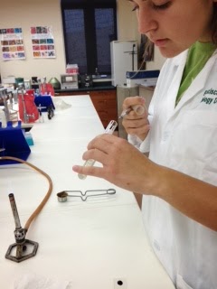
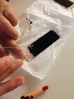
First, Juliet fixed Bacteria J to a clean slide, and then placed it on a staining rack over a beaker of boiling water.  The steam from the water softens the waxes in the cell wall.  To keep the stain in place (the steam could make it run too much) Sierra placed Bilbous paper on top of the slide and then saturated the entire slide with carbol fuchsin.

Sierra and Juliet kept Matt in line with not letting
the stain get dry...he gets distracted so easily!


                            It was very important during this part of the procedure to keep the slide and paper constantly flooded with carbol fuchsin, so the whole cell could be thoroughly stained.  The slide was heated and colored for 5 minutes....


...and then removed and allowed to cool over the sink.   The cooling process is what 'fastens' the stain into the cell wall because during this time the waxes solidify, holding the stain in place.  Matt rinsed the slide with DiH2O... 


...and then rinsed it again with acid alcohol until the color ran clear.  This is a decolorizing agent that removes any stain not held in place by the wax in the cell wall.  If the specimen is a mycobacterium, the
Rinsing slide with decolorizing agent
stain will be held in place and not removed by the decolorizing agent.   


Flooding with methylene blue


The next step allows us to see the bacteria itself.  Matt flooded the slide with methylene blue, allowed it to rest for two minutes and then rinsed the whole slide with DiH20.  After blotting gently with Bilbous paper, our slide was ready for viewing.  So has Dr. P been exposing us to dangerous bacteria again??
DiH2O rinse

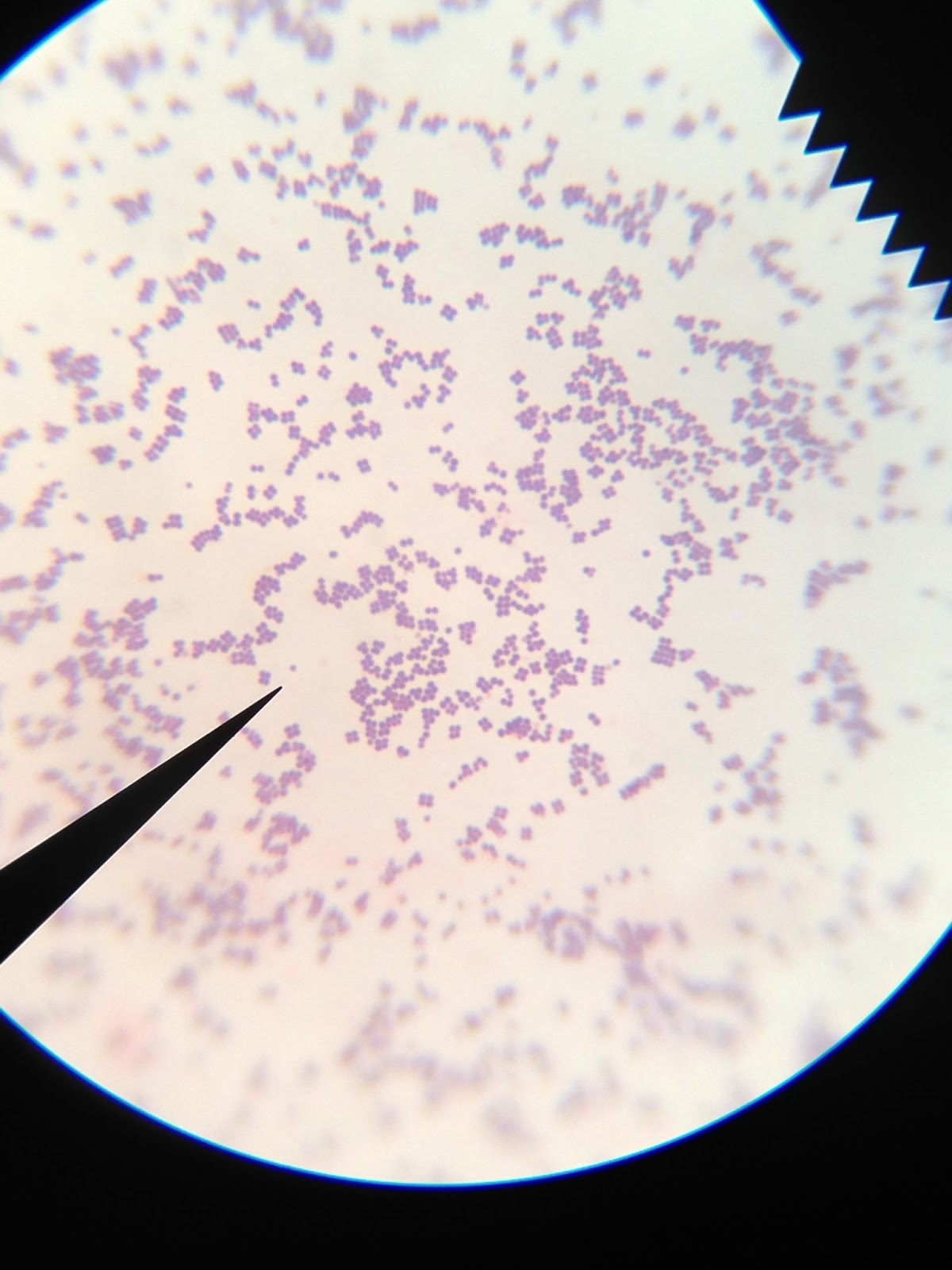
Nah. Dr. P would never do that!  There is no red or pink color whatsoever, which would be visible if these bacteria were a mycobacterium species.  Only blue as far as the eye can see, which comes from the methylene blue stain used.   Phew! 


Next task to learn more about Bacteria J: endospore stain. The stain used is Malachite green. Like the acid-fast stain, heat is used to permeate stain the into the endospore itself.  The thick protein coat  of the endospore does not allow simply staining to take hold, which is why heat and a different stain are necessary, but the protein also holds the stain in the endospore through a rinse of water and safranin.  

The procedure itself is very similar to acid-fast staining, except the slide is heated over steaming water for a few minutes longer (5-6 mins total to be exact).  Here's a quick picture recap: 

Slide over troubled boiling water.  Matt is flooding it with
Malachite green stain.

A precarious removal.  We thought the whole thing was going
to end up in the water.  Matt is going to be watched carefully from
now on.

After cooling, the slide is rinsed with DiH2O. 

 Next, the slide is covered in safranin stain, which then sits for about a minute and a half.  The safranin stains the rest of the bacteria, but not the endospore, which will remain green.  If endospores are present, the slide should look very Christmasy under the microscope.


Rinsing safranin from slide
Look at that beautiful slide. 

The last few steps are pretty standard for us by now; rinse the slide with DiH2O for 30 seconds, blot gently, and appreciate our fine work.   

Blotting gently

So is it Christmas come early in the microbio lab? 
No, not for this group. Although our stain is looking mighty fine as it is.  Good work team.  


 Final order of random business: Dr. P gave us a bacteriophage last week, which we put on a spread plate with Bacteria J.  This was to demonstrate how bacteriophages kill any bacteria  they touch.  It ever-so-sightly prevented bacterial growth in the pattern that we applied the phage, but not by much (the pattern was supposed to be our initials, JSM).  

Tuesday, September 24, 2013

Capsule Stain of Mystery Bacteria


CAPSULE STAIN OF MYSTERY BACTERIA

Class: Tuesday, September 24th, 2013
In today's lab, we prepared our mystery bacteria with a capsule stain. The capsule stain is a type of negative staining. Similar to the negative stain we did in another lab, the purpose of this type of stain is to view bacterial capsules, which are difficult if possible with basic dyes. A bacterial capsule is one type of glycocalyx, the other type is a slime layer. However, slime layers are highly unorganized and easily detach from the cell wall, so the chances of even seeing a slime layer is slim if at all. Therefore, our main focus in this lab is to determine whether our bacteria has a capsule surrounding the cell wall.  If our bacteria has a capsule, a halo surrounding it will remain unstained. This "halo" is the bacterial capsule. 

A little background on bacterial capsules, they consist of polysaccharides usually combined with lipids and proteins, which adds to the organized structure of the capsule. The capsule acts as armor on the bacteria, protecting it from being eaten by phagocytic white blood cells. So bacteria with capsules stand a better chance of surviving in the human body than other bacteria that may not have a capsule.

To start the procedure, we prepared a negative stain of our mystery bacteria. We used the same technique as we did in our previous negative staining lab with nigrosin.


image.jpeg
Negative stain of mystery bacteria using nigrosin


image.jpeg
Magnified view of negative stain bacteria

After we placed our bacteria in a negative stain and let it air dry, we covered it with crystal violet. After the appropriate time of letting the smear soak in crystal violet, we gently rinsed off the excess stain. We were very careful with this step because the smear could very easily be removed. The goal is to NOT remove much of the smear.
image.jpeg

image.jpegimage.jpeg


We blotted the slide dry with pieces of bibulous paper, and then looked at our slide under a microscope to see some pretty cool stuff. However, sadly we saw no capsule surrounding our mystery bacteria...But we took a pretty awesome picture of it!

                                    image.jpeg

Random Dr. P's Experiment: We were given a bacteriophage to be tested with our mystery bacteria J. We inoculated bacteria J and streaked it onto the agar plate in a zig-zag fasion. The bacteriophage was streaked over top in the shape of our initials-JSM. This is to demonstrate how bacteriophage will kill the bacteria it touches.

image.jpeg                image.jpeg                       image.jpeg
                       

Friday, September 20, 2013

More results, Gram Staining an Unknown Bacteria, and That Nasty Tea

More results, gram staining an unknown bacteria, and that nasty tea

Class: Tuesday, September 17, 2013 

Bit of a disappointment today...turns out our bacteria from the air pure culture does not have any magical antibiotic super powers.  All three lines of bacteria grew together over the weekend. 


Both of the white bacteria samples have much wider growth patterns than the orange bacteria, but this is not enough to indicated any antibiotic action going on.  

Last week, Dr. P. gave us an unknown bacterial sample on an agar slant that we will be performing various tests and stains on to observe them under the microscope better.  Our sample is J, to be kept at 25 C. The mystery bacteria is a pale orange color and is shiny, possibly indicating the presence of a capsule or slime layer.  


According to the diagram in the book, our sample grew in a filiform (or even) growth pattern.  It did not spread more than the pattern it was applied with, but filled that line in more fully.  


Our first task today was to transfer some of Bacteria J to a new agar slant which we will grow until Thursday and then store in the fridge for later, in case anything should happen to the current sample.  Juliet was sure to use proper sterile technique and also employed the nifty test-tube-holding-techniques while transferring culture from one test tube to another: 


Double fisting the test tubes: Yes Jess, it is really that awesome
Sterilize by fire! 


Next order of business: Gram stain Bacteria J.  We used the technique last week and are basically experts at this point.  Just a quick recap:

1. Smear bacteria in a drop of water and fix using flame
2. Flood slide with crystal violet and rinse with water after 1 minute
3. Flood slide with Gram's iodine and rinse after 1 minute
4. Use 95% ethanol to decolorize slide
5. Flood smear with safranin color and rinse with water after 1 minute

 Our results? 
Mystery Bacteria J under microscope after
Gram staining
Beautiful! The bacteria took the blue/purple coloring, meaning we know that Bacteria J is Gram positive.  We can also see the shape of the bacteria; they are circular and seem to occur in quads, or little groupings of four bacteria together.


It's tea time! Last week we took a sample from the tea Juliet brought in and made both a streak plate and broth sample of it.  The results are quite intriguing and must be further pursued.  

The streak plate is not particularly exciting, in terms of bacterial growth, but the broth culture has a little more to look at.
Dr. P. was most intrigued that there seems to be some anaerobic growth going on here.  That's why the streak plate has very little, but the broth culture as some very healthy fuzzy bacteria in it, growing in the culture medium, not on top of it.  


In order to gain more understanding, we must of course test the bacteria's ability to grow even more.  We took a sample of the bacterial growing on the streak plate and put it into a broth culture.  We then took a sample of the fuzzy bacteria growing in the broth culture and put it on a streak plate.  I hope our arrows don't get too confusing here!

Tea bacteria sample moved from the broth
to an Petri dish
Tea bacteria sample moved from plate to the broth 


Thursday, September 19, 2013

Negative Stain of Mystery Bacteria & Steubenville Air Sample

Negative Stain of Mystery Bacteria & Steubenville Air Sample

Class: Thursday, September 19, 2013


Today, after checking out the growth our mystery bacteria, we decided to do a negative stain of it.  A negative stain is when you create a dark background around the transparent cells.  The purpose of this type of stain is to see the outline of the bacteria more clearly.


Nigrosin on glass slide

We started by adding a drop of a dark stain called nigrosin on one end of a glass slide.


Next, after sterilizing the innoculating loop, we transfered a small amount of our mystery bacteria from the test tube and mixed in with the drop of nigrosin stain on the glass slide.

Capturing small loop of mystery bacteria

Mixing mystery bacteria into stain


After again sterilizing the innoculating loop, we spread the stain across the glass slide using another clean glass slide, leaving a feathered edge on one end.

Sliding stain across


Feathered edge of stain of mystery bacteria
Magnified view of mystery bacteria


We allowed the stain to air dry, and afterwards we examined the mystery bacteria under a microscope.


Next, we did another negative stain, using the same technique, but this time of the Steubenville air sample:

Capturing small loop of air sample

Sliding stain across
After letting the stain air dry, this is what we viewed under the microscope:

Magnified view of air sample

 Lastly, here is an update on what grew on the agar plate and in the broth from Juliet's tea.  The original growths of the tea were swapped: what grew on the agar plate was swabbed and placed in the broth, and what grew in the broth was swabbed and placed on the agar plate.

Tea bacterial growth