Negative Stain of Mystery Bacteria & Steubenville Air Sample
Class: Thursday, September 19, 2013
Today, after checking out the growth our mystery bacteria, we decided to do a negative stain of it. A negative stain is when you create a dark background around the transparent cells. The purpose of this type of stain is to see the outline of the bacteria more clearly.
 |
| Nigrosin on glass slide |
We started by adding a drop of a dark stain called nigrosin on one end of a glass slide.
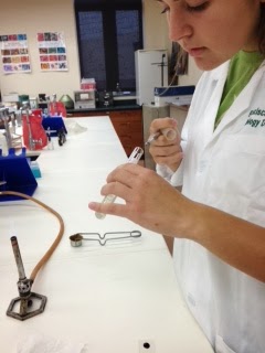
Next, after sterilizing the innoculating loop, we transfered a small amount of our mystery bacteria from the test tube and mixed in with the drop of nigrosin stain on the glass slide.
 |
| Capturing small loop of mystery bacteria |
 |
| Mixing mystery bacteria into stain |
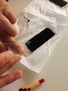
After again sterilizing the innoculating loop, we spread the stain across the glass slide using another clean glass slide, leaving a feathered edge on one end.
 |
| Sliding stain across |
 |
| Sliding stain across |
 |
| Feathered edge of stain of mystery bacteria |
 |
| Magnified view of mystery bacteria |
We allowed the stain to air dry, and afterwards we examined the mystery bacteria under a microscope.
Next, we did another negative stain, using the same technique, but this time of the Steubenville air sample:
 |
| Capturing small loop of air sample |
 |
| Sliding stain across |
After letting the stain air dry, this is what we viewed under the microscope:
 |
| Magnified view of air sample |
Lastly, here is an update on what grew on the agar plate and in the broth from Juliet's tea. The original growths of the tea were swapped: what grew on the agar plate was swabbed and placed in the broth, and what grew in the broth was swabbed and placed on the agar plate.
 |
| Tea bacterial growth |


No comments:
Post a Comment